


Canine Mitral Valve Degenerative Disease (Part 2)
01. Overview
02. Clinical Examination
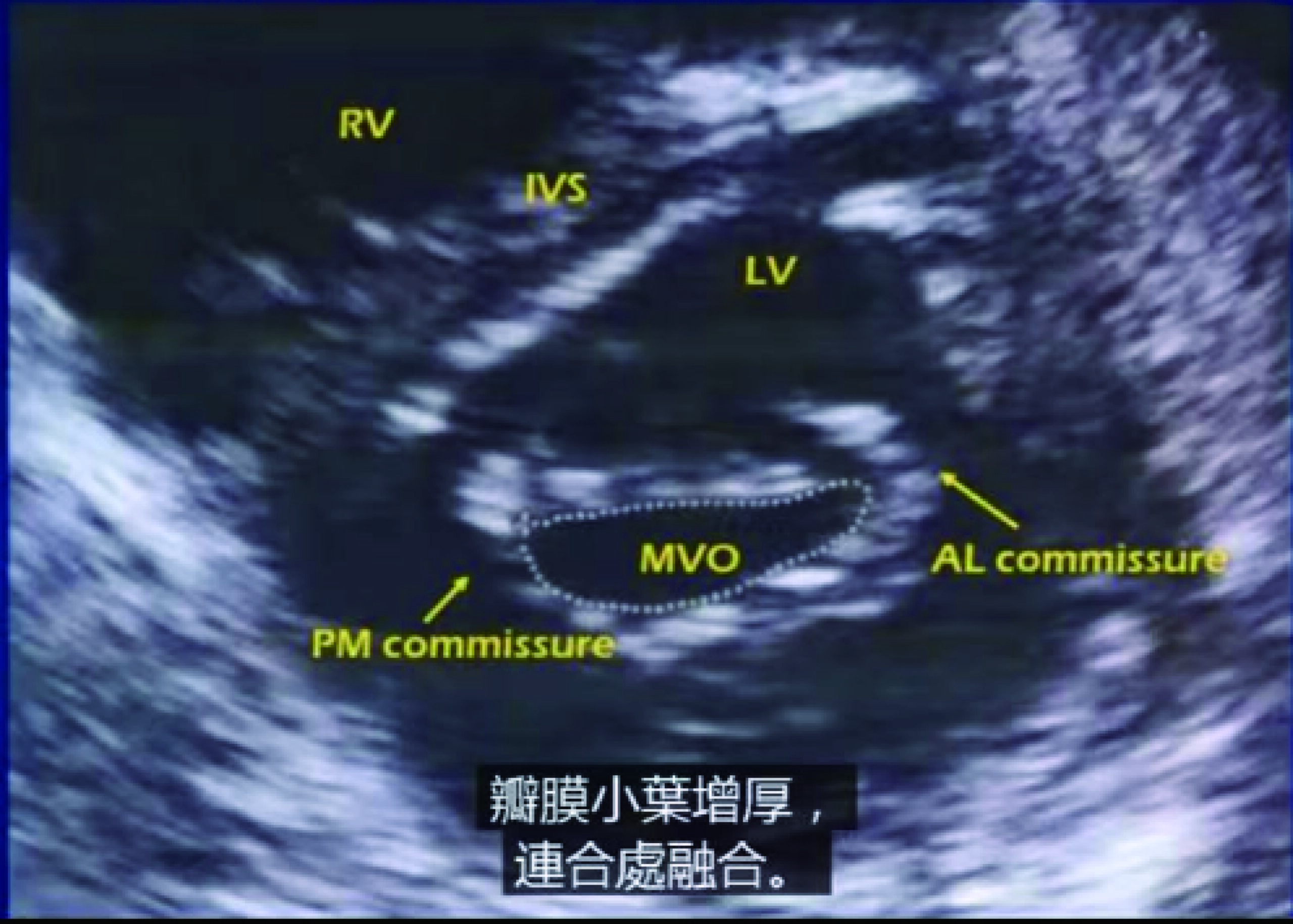
1. Mitral Stenosis (MS)
- Left Atrial Compensation Stage (Early Phase)
- Left Atrial Decompensation Stage (Middle Phase)
Persistent pulmonary congestion (leading to pulmonary hypertension) progresses to right ventricular dilatation, accompanied by ascites and limb edema. At this stage, comprehensive assessment of cardiac chamber dimensions and hemodynamic parameters via BPU60C ultrasound is recommended for treatment planning.
- Correlation with Staging
- Echocardiographic Findings
- Electrocardiographic Features
“Mitral P wave”: A P-wave duration > 0.05 seconds indicates left atrial enlargement.
QRS complex axis deviation to the right; the main waves in leads Ⅰ and Ⅱ show a “peak-to-peak” pattern, suggesting right ventricular enlargement.
Atrial fibrillation may occur in severe cases (to be confirmed by combined echocardiographic assessment with PT50 ultrasound).
2. Mitral Regurgitation (MR)
Pathophysiological Process
Mitral regurgitation → left atrial and left ventricular hypertrophy → left heart failure → right heart failure.
Echocardiographic Findings
Recommended ultrasound devices: BPU60C, PT50.
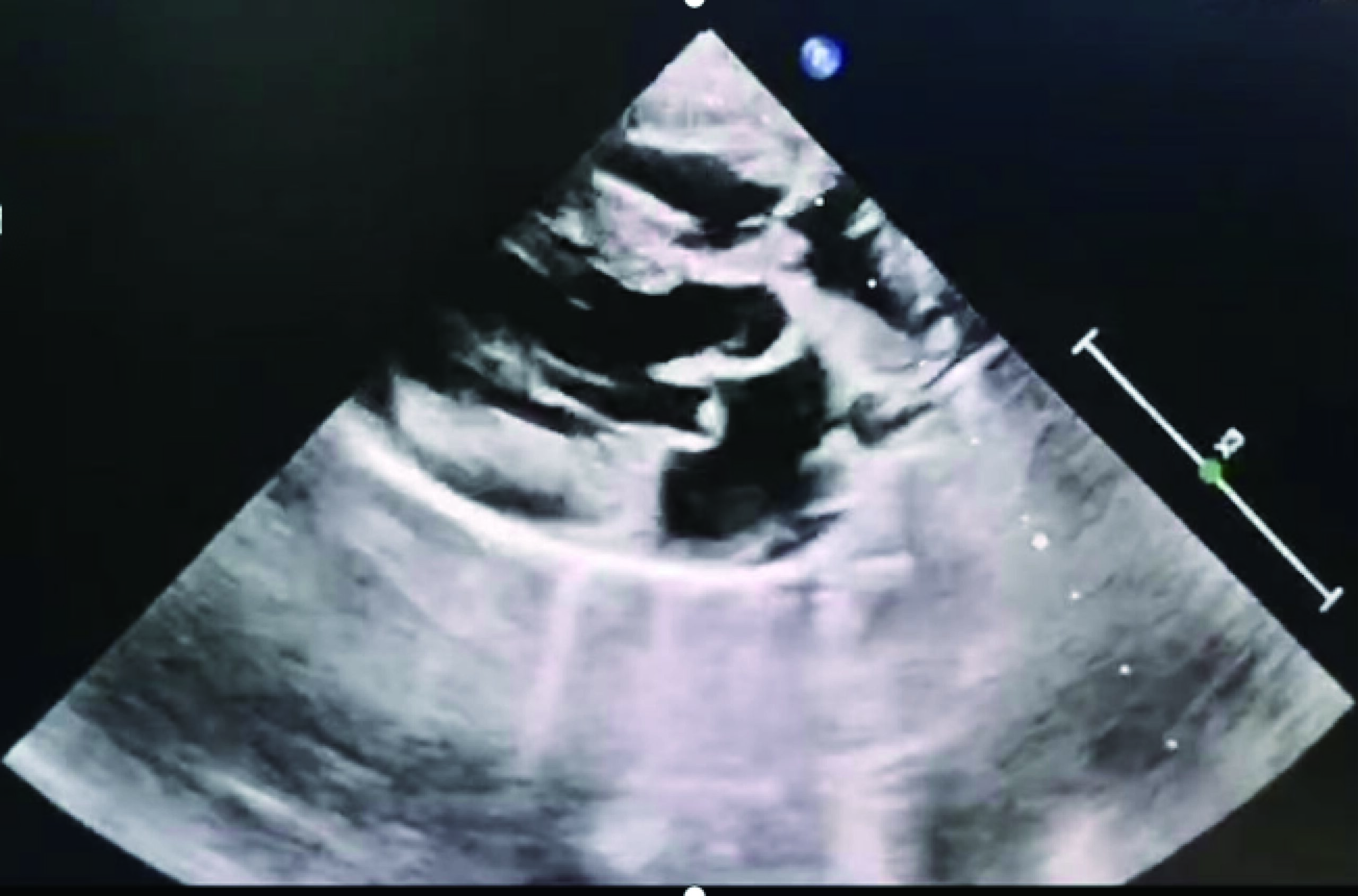
Two-dimensional ultrasound: Mitral leaflet thickening, chordae tendineae thickening, no significant restriction of anterior mitral leaflet movement, increased echo intensity, and valvular prolapse; obvious regurgitation can be observed in some cases.
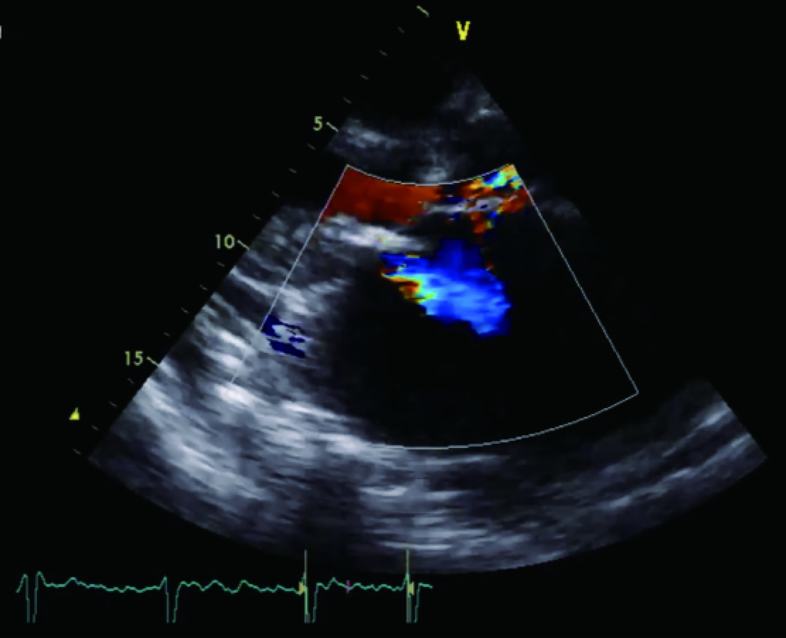
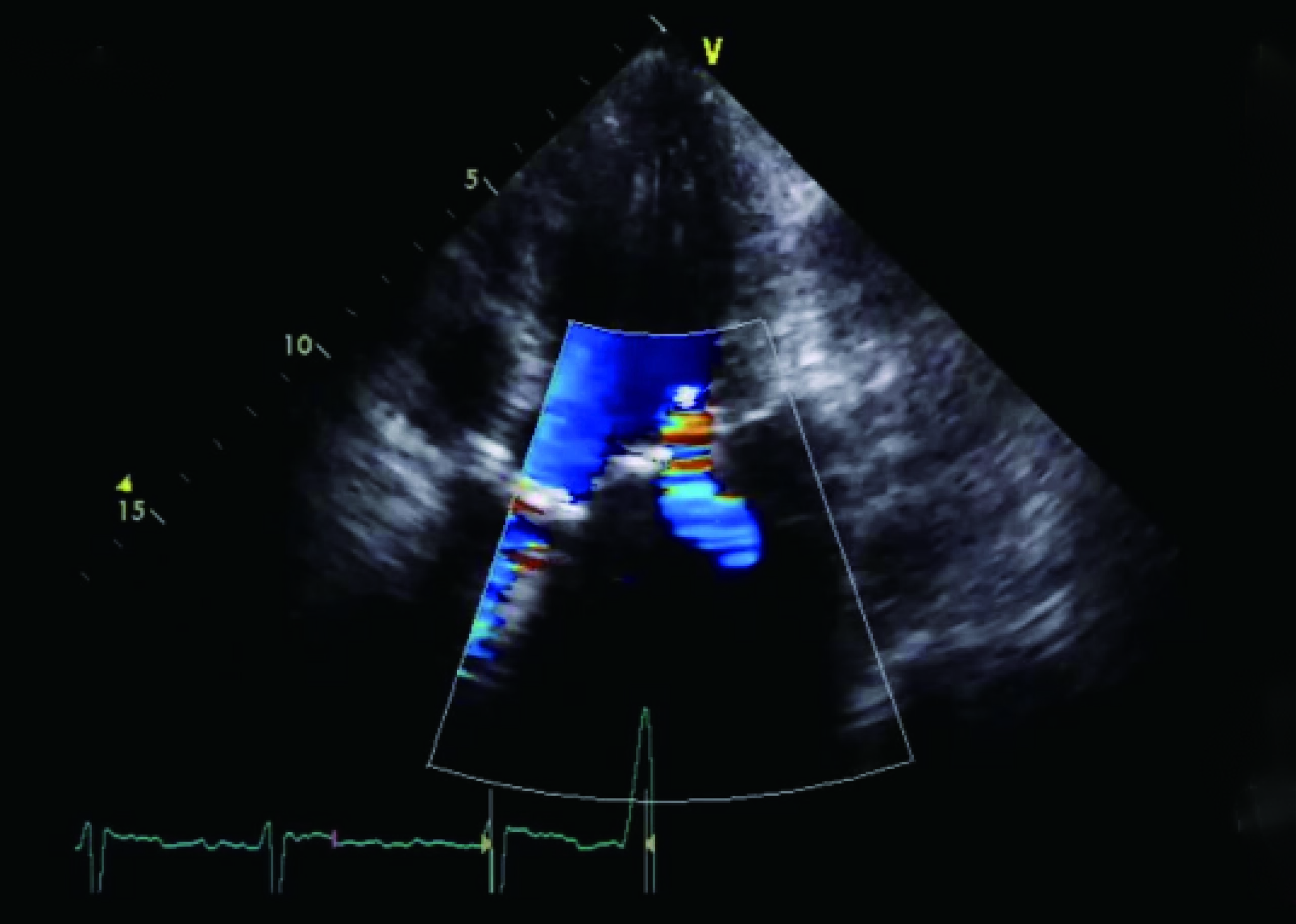
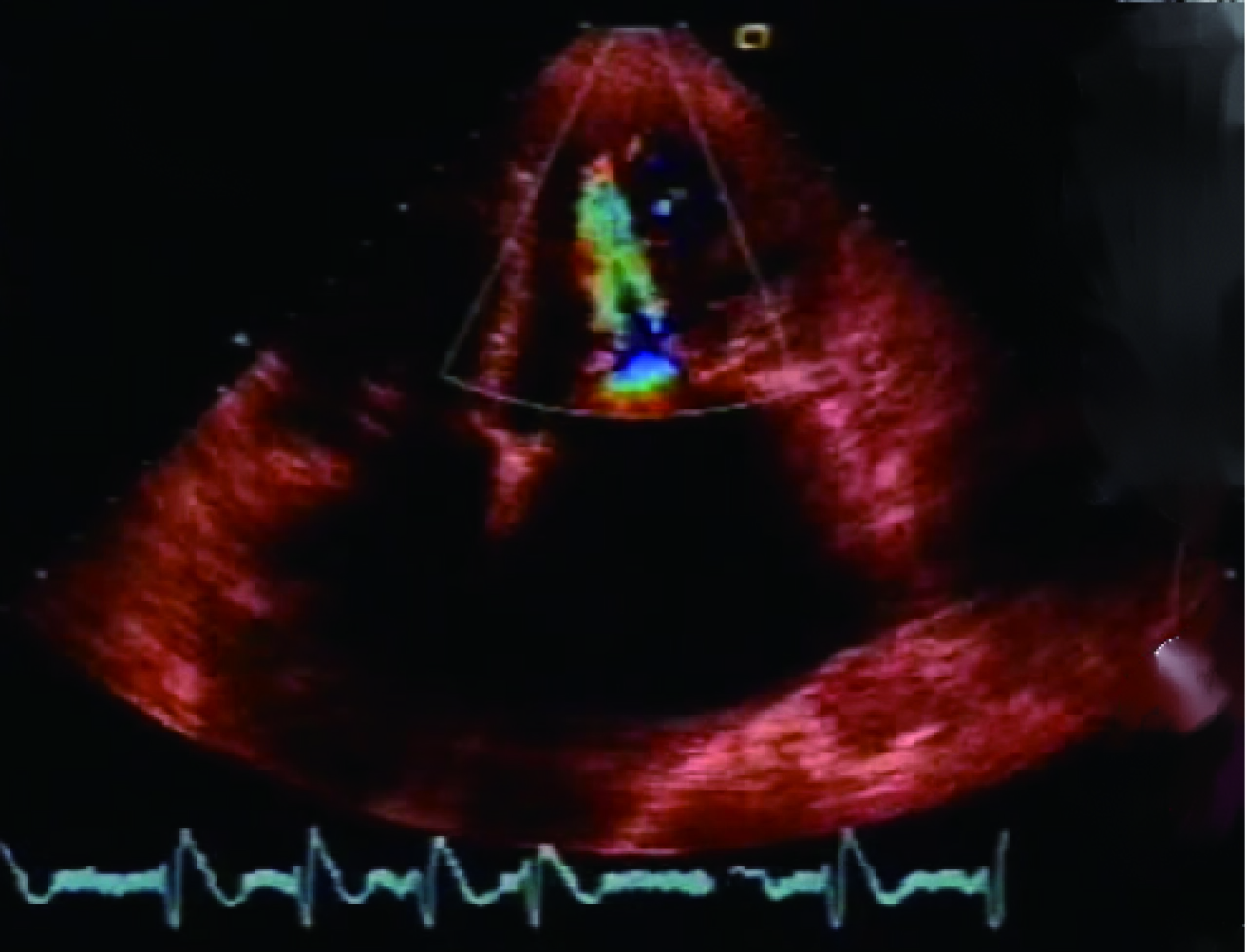
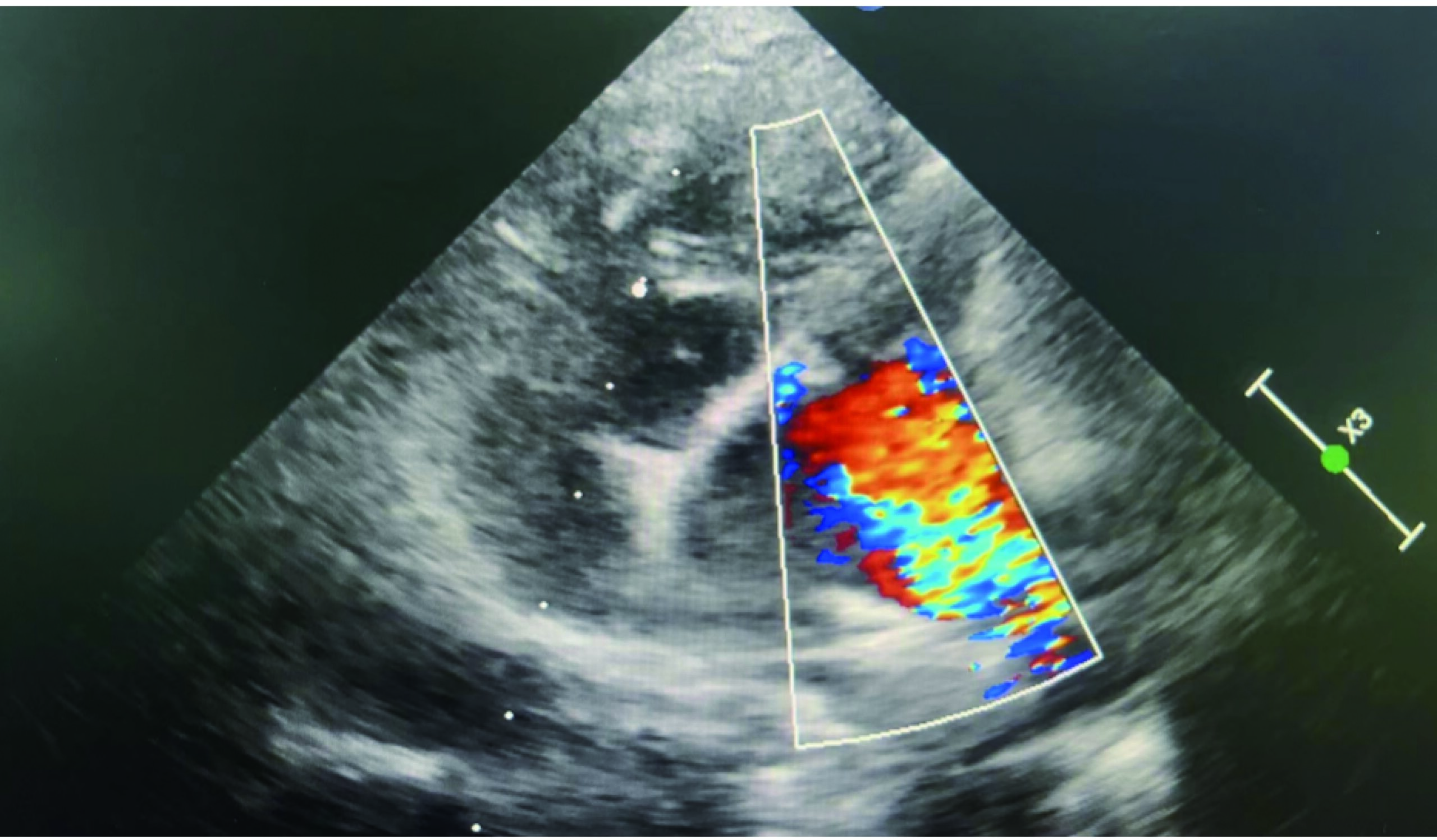
Color Doppler: The gold standard for detecting regurgitation; a multicolored (predominantly blue) regurgitant jet flows from the mitral valve defect into the left atrium during systole. The severity of regurgitation can be assessed by comparison with the left atrial size.
Electrocardiographic Features
Sinus P-wave widening with a biphasic pattern (P-wave duration > 0.05 seconds) indicates left atrial enlargement.
QRS complex axis deviation to the left; main waves in leads Ⅰ and Ⅱ show an “opposite-to-opposite” pattern, suggesting left ventricular enlargement.
Atrial fibrillation may occur in severe cases.
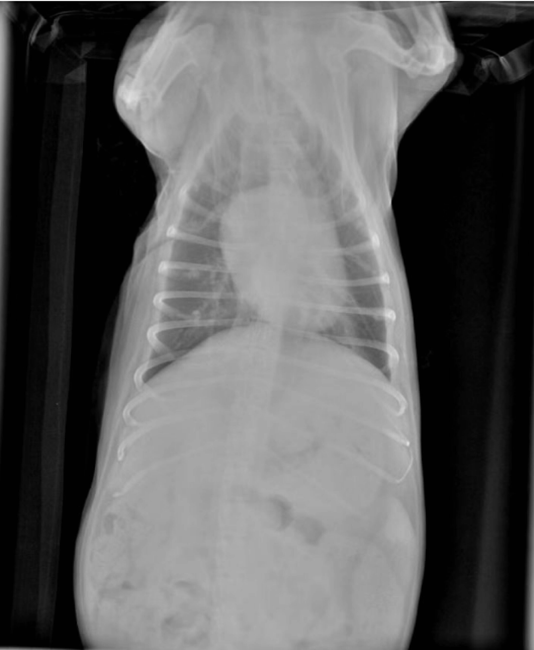
Radiographic Findings
“Spherical” heart.
3. Mitral Stenosis Combined with Regurgitation
Etiology
Infective endocarditis.
Congenital heart disease, etc.
Treatment Principles
Infective cases: Antibiotic therapy (e.g., ceftriaxone sodium, penicillin sodium) is recommended.
Congenital cases: Targeted medication is administered in the absence of surgical intervention (specific treatment methods will be introduced in subsequent articles).
Echocardiographic Findings
Combined manifestations of mitral regurgitation and mitral stenosis. For infective cases, common findings include leaflet, chordae tendineae, and papillary muscle deformation, as well as valvular vegetations (detectable via BPU50 ultrasound for precise lesion localization).